Structure of the human eye
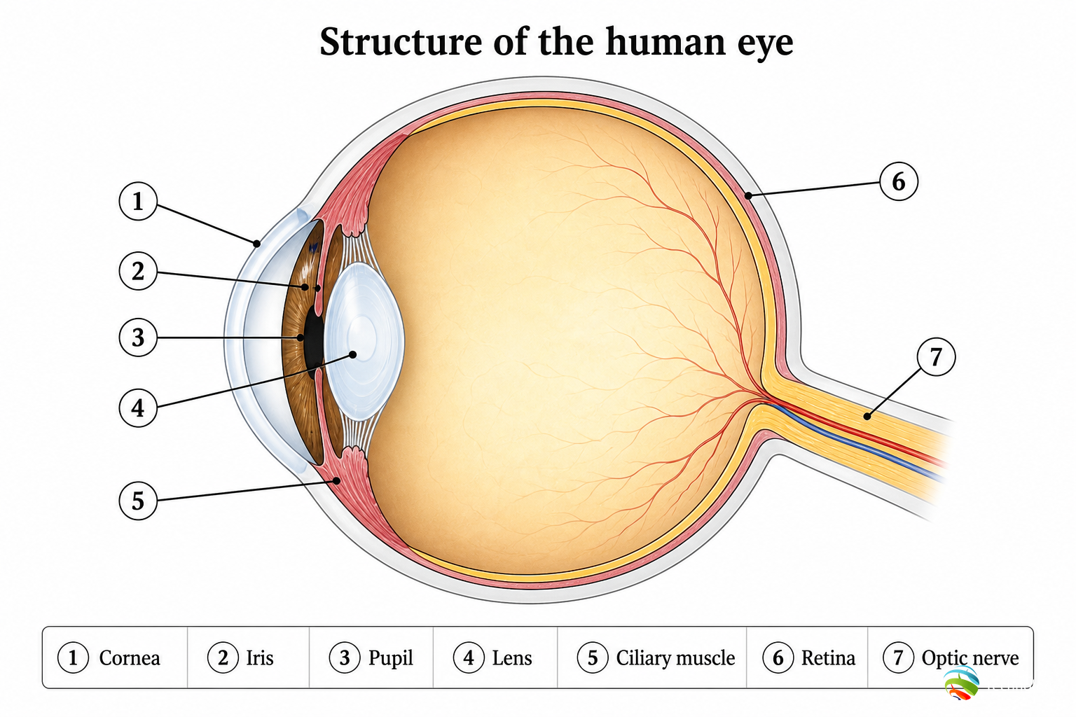
Cornea, iris, pupil, lens, ciliary muscles, retina, optic nerve.
Step 1 — Study the figure
Cover the label key on the image, name each numbered part, then check the explanations below.

Step 2 — Diagram details for students
What this diagram shows
The human eye forms a real inverted image on the retina. Most refraction happens at the cornea, while the lens fine-tunes focus by accommodation. Iris controls light entry, and optic nerve carries signals to the brain.
Numbered parts — mark & remember
Match each number on the figure with the meaning below. Say the function aloud once.
1 Cornea
The cornea is the transparent front covering and provides most of the eye’s refraction. Light bends strongly here before reaching the lens. Injury to cornea seriously affects clear vision.
2 Iris / pupil
The iris is the coloured diaphragm that controls pupil size. In bright light the pupil becomes smaller; in dim light it widens. This regulates how much light reaches the retina.
3 Eye lens & ciliary muscles
The eye lens provides fine focusing. Ciliary muscles change lens thickness for accommodation between near and far objects. Near point is about 25 cm for a normal adult eye.
4 Retina
The retina is the light-sensitive screen where the image forms. It contains cells that send signals toward the optic nerve. A real inverted image is formed here in normal vision.
5 Optic nerve
The optic nerve carries visual information from retina to brain. The brain interprets signals as upright meaningful sight. The blind spot lacks receptor cells where the nerve leaves.
How to read this figure
- Cornea does major refraction; lens does accommodation.
- Image on retina is real and inverted.
- Near point ~25 cm; far point at infinity for normal eye.
Redraw for board marks
- Draw side section of eyeball.
- Label cornea, iris/pupil, lens, retina, optic nerve.
- Mark ciliary muscles near the lens.
Board answer tip: Draw eye section with seven labels and write one function each for cornea, lens, and retina.
Exam tips — don’t lose marks
- Accommodation is focal-length change by ciliary muscles.
- Most refraction is at cornea, not only lens.
- Blind spot has no photoreceptors.