Leaf cross-section
Epidermis, mesophyll with chloroplasts, stomata, xylem and phloem.
Step 1 — Study the figure
Cover the label key on the image, name each numbered part, then check the explanations below.

Step 2 — Diagram details for students
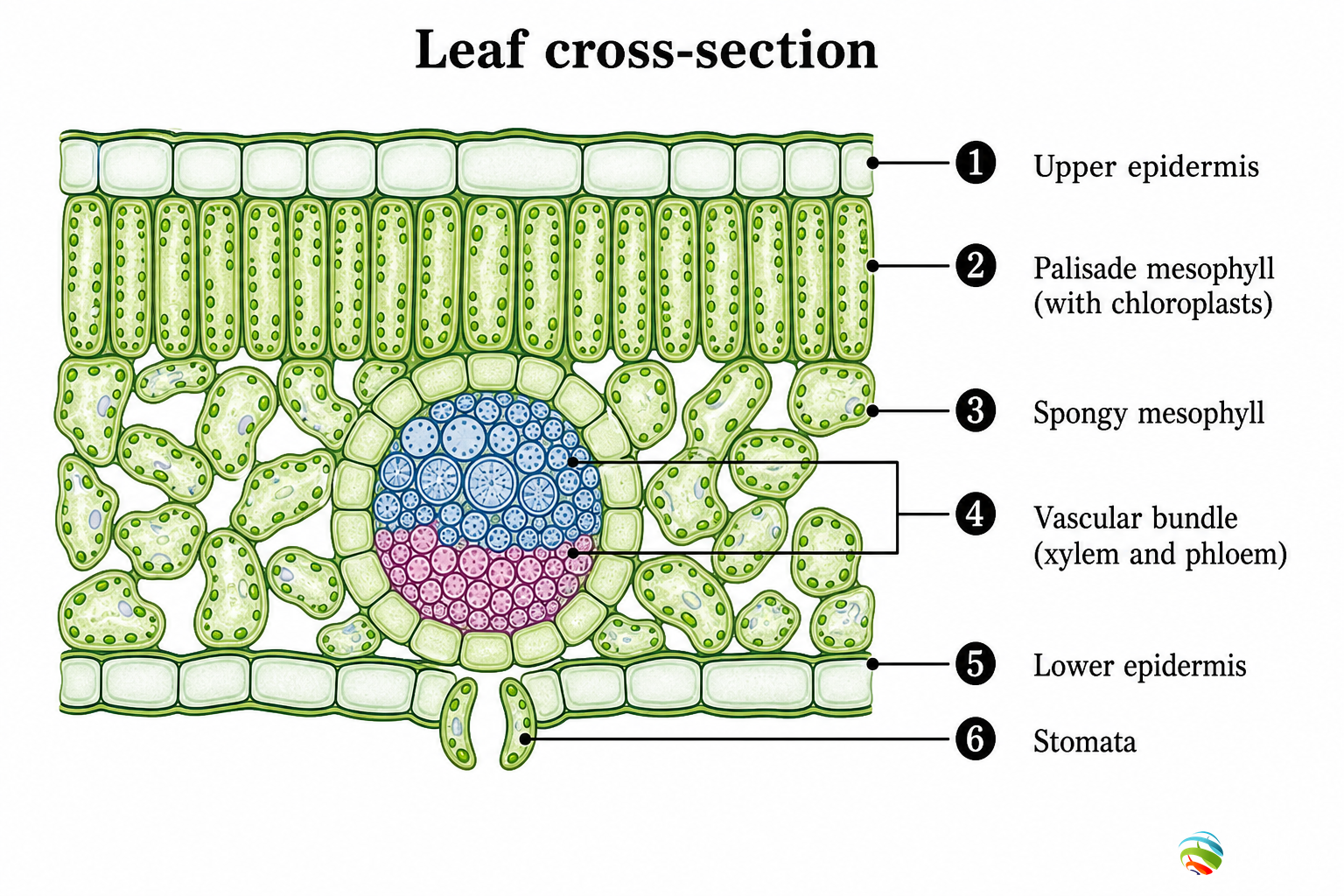
What this diagram shows
A leaf cross-section shows how the leaf is built for photosynthesis and gas exchange. Palisade cells capture light, air spaces help gases move, stomata control exchange, and veins transport water and food. Label layers in order from upper to lower surface in exams.
Numbered parts — mark & remember
Match each number on the figure with the meaning below. Say the function aloud once.
1 Upper epidermis
This is a protective layer of cells on the top surface of the leaf. It is often covered by a waxy cuticle that reduces unwanted water loss. It allows light to pass through to the photosynthetic cells below.
2 Palisade mesophyll
These are elongated, closely packed cells rich in chloroplasts. They are the main site of photosynthesis because they receive most light. In diagrams, show many green chloroplasts inside these cells.
3 Spongy mesophyll
These cells are loosely arranged with large intercellular air spaces. The spaces allow carbon dioxide and oxygen to diffuse to and from cells. They also contain some chloroplasts, but fewer than palisade tissue.
4 Vascular bundle
The vein contains xylem and phloem. Xylem brings water and minerals from roots to the leaf. Phloem exports sugars made in photosynthesis to other parts of the plant.
5 Lower epidermis
This protective layer covers the underside of the leaf. In many plants it contains more stomata than the upper epidermis. It helps prevent damage while still allowing controlled gas exchange.
6 Stomata
Stomata are pores bordered by guard cells. They let CO₂ enter for photosynthesis and let O₂ and water vapour leave. Guard cells open or close the pore by becoming turgid or flaccid.
How to read this figure
- Read the leaf from top to bottom: epidermis → palisade → spongy → lower epidermis.
- Connect structure to function: chloroplasts for food making, stomata for gases, veins for transport.
- Mention the open/close stomata trade-off with transpiration.
Redraw for board marks
- Draw layered rectangles for epidermis and mesophyll.
- Add a round vascular bundle with xylem and phloem regions.
- Show one stoma with two guard cells on the lower side.
- Number parts and write a neat key.
Board answer tip: Draw the layered section, number six parts, and write one function for each label in the key.
Exam tips — don’t lose marks
- Always label chloroplasts in palisade if photosynthesis is the focus.
- Do not forget xylem vs phloem roles in the vein.
- Stomata answers should include guard cells and turgidity.